Zygomycota
Zygomycota, or zygote fungi, is a former division or phylum of the kingdom Fungi. The members are now part of two phyla the Mucoromycota and Zoopagomycota.[1] Approximately 1060 species are known.[2] They are mostly terrestrial in habitat, living in soil or on decaying plant or animal material. Some are parasites of plants, insects, and small animals, while others form symbiotic relationships with plants.[3] Zygomycete hyphae may be coenocytic, forming septa only where gametes are formed or to wall off dead hyphae. Zygomycota is no longer recognised as it was not believed to be truly monophyletic.[1]
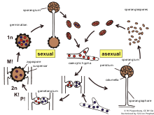
The name Zygomycota refers to the zygosporangia characteristically formed by the members of this clade, in which resistant spherical spores are formed during sexual reproduction. Zygos is Greek for "joining" or "a yoke", referring to the fusion of two hyphal strands which produces these spores, and -mycota is a suffix referring to a division of fungi.[4]
The term "spore" is used to describe a structure related to propagation and dispersal. Zygomycete spores can be formed through both sexual and asexual means. Before germination the spore is in a dormant state. During this period, the metabolic rate is very low and it may last from a few hours to many years. There are two types of dormancy. The exogenous dormancy is controlled by environmental factors such as temperature or nutrient availability. The endogenous or constitutive dormancy depends on characteristics of the spore itself; for example, metabolic features. In this type of dormancy, germination may be prevented even if the environmental conditions favor growth.
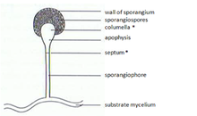
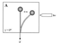
In zygomycetes, mitospores (sporangiospores) are formed asexually. They are formed in specialized structures, the mitosporangia (sporangia) that contain few to several thousand of spores, depending on the species. Mitosporangia are carried by specialized hyphae, the mitosporangiophores (sporangiophores). These specialized hyphae usually show negative gravitropism and positive phototropism allowing good spore dispersal. The sporangia wall is thin and is easily destroyed by mechanical stimuli (e.g. falling raindrops, passing animals), leading to the dispersal of the ripe mitospores. The walls of these spores contain sporopollenin in some species. Sporopollenin is formed out of β-carotene and is very resistant to biological and chemical degradation. Zygomycete spores may also be classified in respect to their persistence:
Chlamydospores are asexual spores different from sporangiospores. The primary function of chlamydospores is the persistence of the mycelium and they are released when the mycelium degrades. Chlamydospores have no mechanism for dispersal. In zygomycetes the formation of chlamydospores is usually intercalar. However, it may also be terminal. In accordance with their function chlamydospores have a thick cell wall and are pigmented.
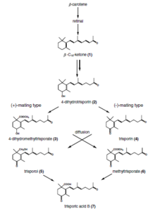
Zygophores are chemotropic aerial hyphae that are the sex organs of Zygomycota, except for Phycomyces in which they are not aerial but found in the substratum. They have two different mating types (+) and (-). The opposite mating types grow towards each other due to volatile pheromones given off by the opposite strand, mainly trisporic acid and its precursors. Once two opposite mating types have made initial contact, they give rise to a zygospore through multiple steps.


