Chytridiomycota
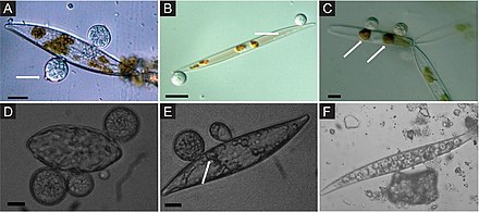
Chytridiomycota are a division of zoösporic organisms in the kingdom Fungi, informally known as chytrids. The name is derived from the Ancient Greek χυτρίδιον (khutrídion), meaning "little pot", describing the structure containing unreleased zoöspores. Chytrids are one of the early diverging fungal lineages, and their membership in kingdom Fungi is demonstrated with chitin cell walls, a posterior whiplash flagellum, absorptive nutrition, use of glycogen as an energy storage compound, and synthesis of lysine by the α-amino adipic acid (AAA) pathway.[2][3]
Chytrids are saprobic, degrading refractory materials such as chitin and keratin, and sometimes act as parasites.[4] There has been a significant increase in the research of chytrids since the discovery of Batrachochytrium dendrobatidis, the causal agent of chytridiomycosis.[5][6]
Species of Chytridiomycota have traditionally been delineated and classified based on development, morphology, substrate, and method of zoöspore discharge.[7][4] However, single spore isolates (or isogenic lines) display a great amount of variation in many of these features; thus, these features cannot be used to reliably classify or identify a species.[7][4][8] Currently, taxonomy in Chytridiomycota is based on molecular data, zoöspore ultrastructure and some aspects of thallus morphology and development.[7][8]
In an older and more restricted sense (not used here), the term "chytrids" referred just to those fungi in the class Chytridiomycetes. Here, the term "chytrid" refers to all members of Chytridiomycota.[2]
The chytrids have also been included among the Protoctista,[7] but are now regularly classed as fungi.
In older classifications, chytrids, except the recently established order Spizellomycetales, were placed in the class Phycomycetes under the subphylum Myxomycophyta of the kingdom Fungi. Previously, they were placed in the Mastigomycotina as the class Chytridiomycetes.[9] The other classes of the Mastigomycotina, the Hyphochytriomycetes and oömycetes, were removed from the fungi to be classified as heterokont pseudofungi.[10]
